
Hijacking Immunity: deciphering how Pancreatic Cancer Driver Cells (PCDCs) orchestrate tumour initiation
Primary supervisor: Beatriz Salvador, Queen Mary University of London
Secondary supervisor: Pilar Acedo, UCL
Project
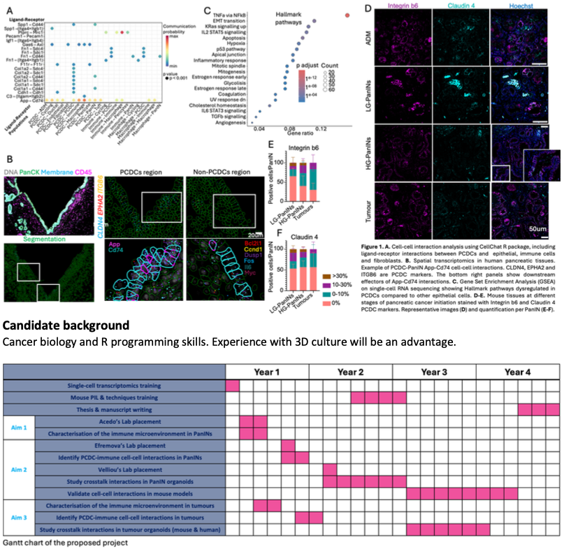
Background: We have previously identified a rare sub-population of epithelial cells called Pancreatic Cancer Driver Cells (PCDCs) that drive pancreatic cancer initiation (manuscript in preparation). PCDCs can interact with their neighbours (Figure1A-B) in early pancreatic pre-malignant lesions (known as PanINs). Interestingly, PCDCs activate different pathways which promote resistance to environmental stressors. Among these pathways are inflammatory signatures including ‘Hallmark_of_inflammatory_response’, ‘Hallmark_Il2-Stat5’ and‘Hallmark_Il6-Stat3’ signalling (Figure1C).
Hypothesis: PCDCs regulate their immune microenvironment during pancreatic cancer initiation, making it more permissible for PanINs to become malignant and develop into pancreatic cancer.
Aims and methods: Here we aim (1) to characterise the immune microenvironment in pancreatic cancer initiation, (2) to study PCDCs interactions with the immune compartment and identify their role in PanIN progression into tumours and (3) to decipher the relevance of PCDC-immune cell-cell interactions in tumours.
Aim 1. Immune microenvironment characterisation: Recent studies show that 60% of healthy adults carry PanINs in their pancreas, however, only ~0.005%/year develop pancreatic cancer, suggesting only some PanINs can progress into tumours. We found that only a few of PanINs contain more than 30% of PCSCs (Figure1D-F). This may suggest that only PanINs with a high proportion of PCDCs can progress into tumours. We will define the immune microenvironment [1,2] surrounding PCDC-rich (PCDChigh) and PCDC-poor (PDCDlow) PanINs to understand if PCDChigh PanINs contain a more permissive immune microenvironment. We will use banked whole genome spatial transcriptomics in mouse [3] tissues (Pdx1CreERT;KRasG12D/+;Rosa26RFP; Figure1B) that recapitulate the different stages of pancreatic cancer initiation and identify PCDChigh and PDCDlow PanINs to define the different immune cell types present in each PanIN type (e.g., macrophages, T cells). We will characterise the immune cell activation state in different PanINs.
Aim 2. PCDC-immune cell-cell interactions in pancreatic cancer initiation: Previous experiments in the lab showed that PCDCs interact with other epithelial cells, fibroblasts and immune cells (Figure1A-B). In this objective we will (a) characterise the interactions between PCDCs and immune cells in PanINs; and (b) understand their role in pancreatic cancer initiation. Using our spatial transcriptomics data and crosstalk analysis R packages (CellChat, CellPhoneDB4), we will identify potential cell-cell interactions. These interactions will be tested in vitro using organoid [5] co-culture systems combining mouse PanIN organoids and immune cells. Finally, interactions will be validated in mouse models.
Aim 3. PCDCs interactions with immune cells in pancreatic cancer: We hypothesise that PCDCs become the Cancer Stem Cells in tumours, a population characterised by their avoidance of the immune system. To understand the crosstalk between PCDCs and immune cells in tumours, we will use whole transcriptome spatial transcriptomics data (as described above) in mouse pancreatic tumours. To confirm our results in human tissues, we will use our banked spatial transcriptomics data from human pancreatic tumours, and human tumour organoid co-cultures with immune cells.
Relevance: Understanding how pancreatic cancer develops can lead to the development of early detection and prevention strategies for high-risk populations as well as unravel novel therapeutic targets.
Potential placements
- Pilar Acedo, Institute for Liver and Digestive Health, UCL
- Eirini Velliou, Department of Targeted Intervention, UCL
- Mirjana Efremova, Barts Cancer Institute, Queen Mary University of London
References
- Fraser, K., Rosado, M., Pereira, S. P. & Acedo, P. Abstract B023: Unravelling fibroinflammatory and immune signatures for pancreatic cancer early detection. Cancer Res 84, B023 (2024).
- Opitz, F. V., Haeberle, L., Daum, A. & Esposito, I. Tumor Microenvironment in Pancreatic Intraepithelial Neoplasia. Cancers 13, 6188 (2021).
- Salvador-Barbero, B., Alatsatianos, M., Morton, J. P., Sansom, O. J. & Hogan, C. KRASG12D Cells Override Homeostatic Cell Elimination Mechanisms in Adult Pancreas Via Wnt5a and Cell Dormancy. Gastroenterology 169, 983-999.e21 (2025).
- Efremova, M., Vento-Tormo, M., Teichmann, S. A. & Vento-Tormo, R. CellPhoneDB: inferring cell–cell communication from combined expression of multi-subunit ligand–receptor complexes. Nat Protoc 15, 1484–1506 (2020).
- Kataki, A.-D. et al. Mapping Tumor–Stroma–ECM Interactions in Spatially Advanced 3D Models of Pancreatic Cancer. ACS Appl. Mater. Interfaces 17, 16708–16724 (2025).