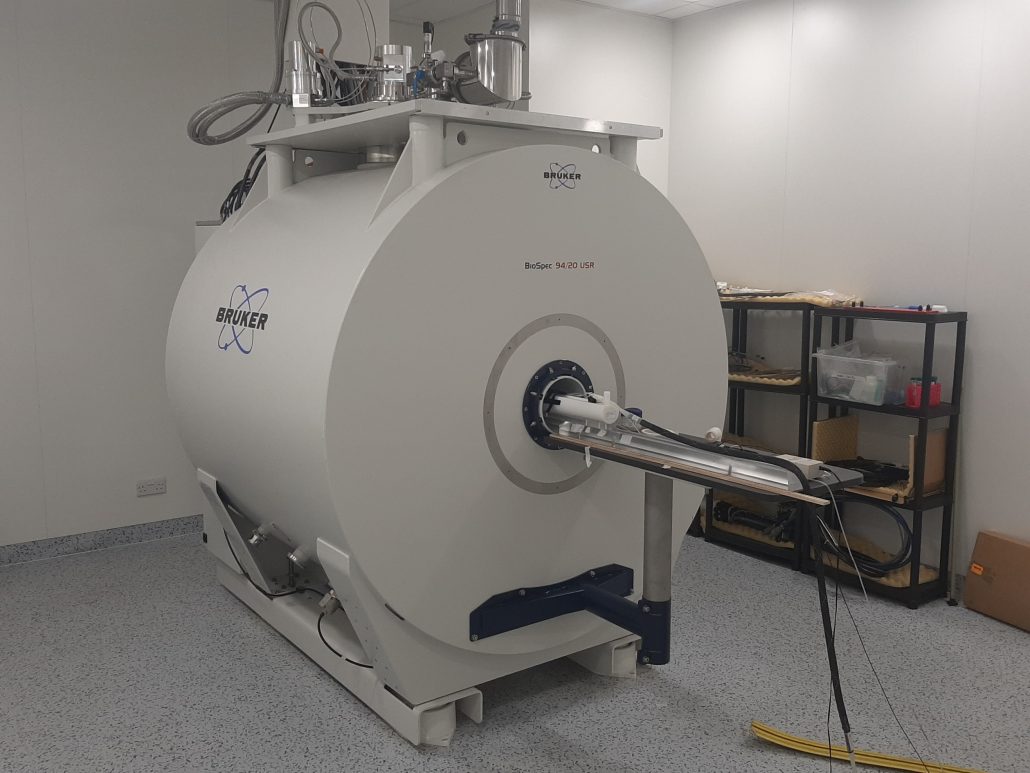
Preclinical 9.4T Magnetic Resonance Imaging (MRI)
The Bruker 9.4T preclinical MRI is based at the UCL Centre for Advanced Biomedical Imaging (CABI). It is equipped with a range of coils designed for investigations in a variety of body regions (e.g. brain, myocardium and abdomen).
The facility provides a range of MRI techniques and post-processing tools to support a range of projects carried out within the CRUK City of London Centre. Thanks to the high magnetic field of this MRI scanner we can track and quantify the impact of biological therapies, thereby increasing our understanding of the tumour microenvironment and enhance the delivery and efficacy of biological therapies. Providing a virtual link between micro and macro-structures, these studies can give new insight into the observed alterations.
Moreover, the MRI is a non-invasive tool that enables longitudinal studies and thus the reduction of animals needed in research studies. The outcome of imaging studies carried out in animal models can suggest targets for intervention in humans. Conversely, findings in human studies can hint at targets for imaging studies in animal models, with the advantages of post-mortem analysis, not feasible in humans.

Contact
Academic Leads
Protocols available
The tumour growth curve can be longitudinally assessed by means of T2 and/or T1 weighted images. [1]
1 – Ramasawmy R. et al. Monitoring the Growth of an Orthotopic Tumour Xenograft Model: Multi-Modal Imaging Assessment with Benchtop MRI (1T), High-Field MRI (9.4T), Ultrasound and Bioluminescence. PLoS One. 2016;11(5):e0156162. Published 2016 May 25. doi:10.1371/journal.pone.0156162
The state-of-the-art diffusion MRI (dMRI) techniques can be adopted to track the evolution of the tumour microenvironment. Water molecule displacement in dMRI-targeted tissues is strongly influenced by the direction, permeability, and number of barriers (cell membranes and myelin sheaths) in the tissue, and by differences in intracellular proteins and organelles. Thus, dMRI can indirectly infer the tissues microstructures. [2]
2 – Roberts, T. A. et al (2020). Noninvasive diffusion magnetic resonance imaging of brain tumour cell size for the early detection of therapeutic response. Sci Rep, 10 (1), 9223. doi:10.1038/s41598-020-65956-4
Thanks to the high strength of its magnetic field, our magnet provides this non-invasive technique to study the biochemical processes characterizing the metabolism of different body regions. [3]
3 – Hulsey KM. et al. ¹H MRS characterization of neurochemical profiles in orthotopic mouse models of human brain tumors. NMR Biomed. 2015;28(1):108-115. doi:10.1002/nbm.3231
This technique is based on the phenomenon of chemical exchange of protons between water and different chemical compounds present in the tissues. Due to their different chemical environment, protons belonging to different compounds resonate at a frequency different to the protons belonging to the water. During signal acquisition, radiofrequency pulses can be transmitted in a frequency range that corresponds to these chemical shifts enabling the selective saturation of these protons. This saturation is transferred from the solute protons to the protons of the water resulting in attenuation of the total water signal. This gives the opportunity to indirectly measure metabolites present in tissues at a small concentration. [4]
4 – Walker-Samuel S. et al. In vivo imaging of glucose uptake and metabolism in tumors. Nat Med. 2013;19(8):1067-1072. doi:10.1038/nm.3252
We are now implementing this technology which enables the quantitative assessing of the tissue mechanical properties such as the stiffness. It has been shown that increased stiffness in the extracellular matrix (ECM) contributes to tumour progression and metastasis. Therefore, MRE can help in the development of stromal modulating therapies and accompanying biomarkers to target ECM stiffness. [5]
5 – Li J. et al. Investigating the Contribution of Collagen to the Tumor Biomechanical Phenotype with Noninvasive Magnetic Resonance Elastography. Cancer Res. 2019;79(22):5874-5883. doi:10.1158/0008-5472.CAN-19-1595


