
First Prize
John Riches (BCI)
A deathly rose
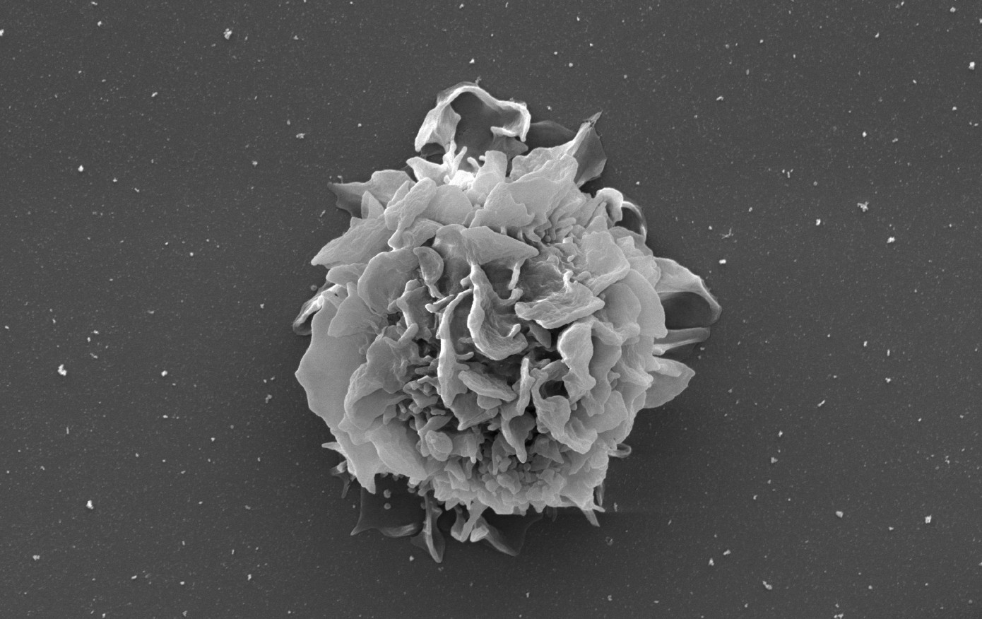
At first glance, this image looks like a delicate flower in bloom. In reality, it is a single leukaemia cell taken from a patient with hairy cell leukaemia, captured using a powerful scanning electron microscope.
The “petals” are tiny projections covering the cell’s surface. These unusual structures give the disease its name and help identify it under a light microscope.
Hairy cell leukaemia is a rare, slow-growing blood cancer that develops in the bone marrow, where blood cells are made. As the cancer cells multiply, they can crowd out healthy blood cells and build up in the blood and spleen, leaving patients tired, vulnerable to infection and living with an enlarged spleen.
Scientists think these hair-like projections may do more than identify the disease. By increasing the cell’s surface area, they may help leukaemia cells interact with and cling to supportive tissues in the bone marrow and spleen, aiding their survival. Research into the genetic changes that cause these projections has also helped scientists understand how hairy cell leukaemia develops, paving the way for important new targeted treatments known as BRAF inhibitors.
This image captures both the disease’s defining feature and the biological insight that helped inspire more effective treatments.
Joint Second Prize
Thomas Willott (BCI)
On the origin of medulloblastoma
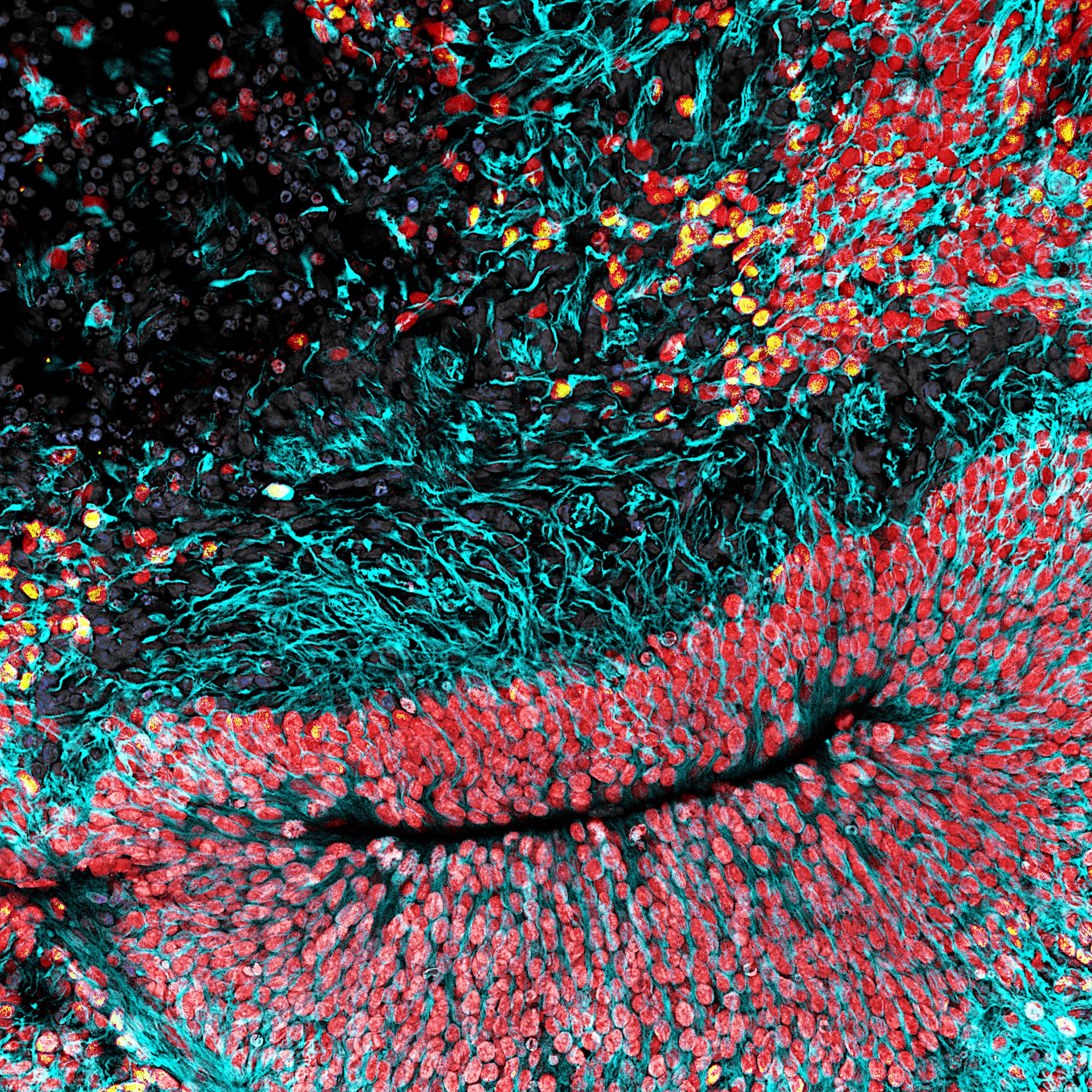
This image offers a glimpse into the earliest stages of human brain development. It shows an organoid – a miniature, lab-grown model that mimics key stages of early brain development, created from stem cells.
The different colours reveal distinct cell populations as they grow and organise themselves. Neural stem cells are shown in red, while turquoise cells represent young developing neurons migrating outwards as the brain takes shape. Nestled among them are rare yellow cells that researchers believe may be the starting point of medulloblastoma, the most common malignant brain tumour in children.
Because these cells exist only during a brief period of foetal development, they are extremely difficult to study directly. Organoids allow researchers to recreate this stage of development in the laboratory and investigate what happens when normal growth goes wrong.
Using this model, Willott and other scientists have shown that disrupting the normal development of these cells can trigger the formation of medulloblastoma. By better understanding how and why these tumours form, researchers hope to identify new cancer pathways which can be targeted, leading to kinder treatment for patients and reducing the need for chemotherapy and radiotherapy which can have severe long-term side effects.
Shahab Aslani (UCL)
Untangling micro-scale vascular networks in lung cancer
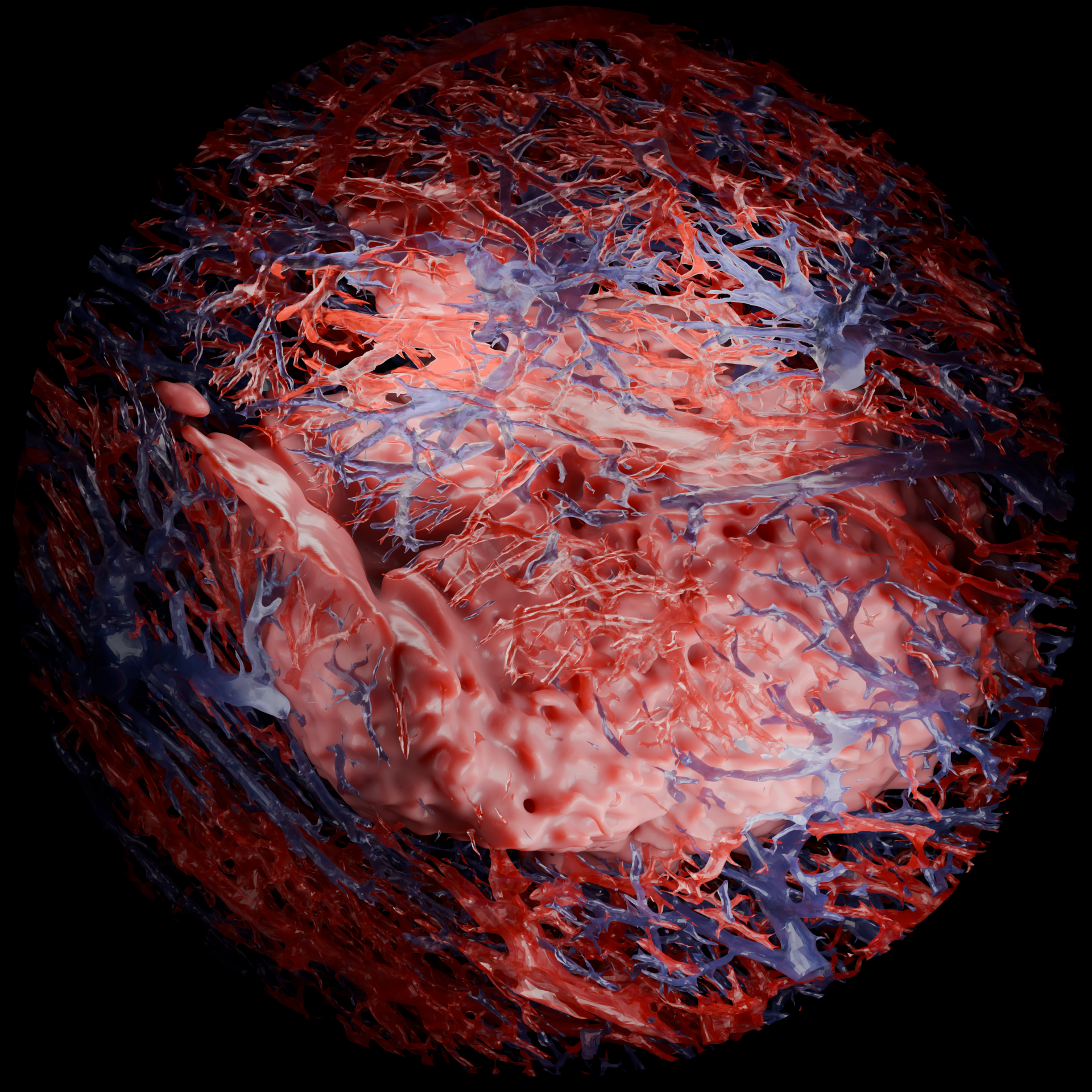
This striking image reveals a hidden three-dimensional world inside a lung tumour. Shown in pink, the 1.6 cm cancer is surrounded by a dense network of blood vessels, with arteries in red and veins in blue.
Like all living tissues, tumours need a blood supply to survive. These vessels deliver oxygen and nutrients that support tumour growth and may facilitate its spread. Understanding how these networks form and develop is therefore an important part of cancer research.
Traditionally, studying the blood vessels inside a tumour has required painstaking reconstruction from hundreds of consecutive stained pathology slides. Here, researchers used hierarchical phase-contrast tomography imaging to visualise the network throughout the tumour and its surrounding tissues in three dimensions, without tissue staining or permanent tissue manipulation.
This detailed map of the tumour’s internal landscape reveals the complex branching patterns of blood vessels and the routes through which blood flows around and within the cancer. Improving our understanding of how lung cancers build and exploit the blood supply they need to grow, could help researchers identify new targets for treatment and develop more effective therapies for people with lung cancer.
George Nicholson (UCL)
The 10 μm galaxy
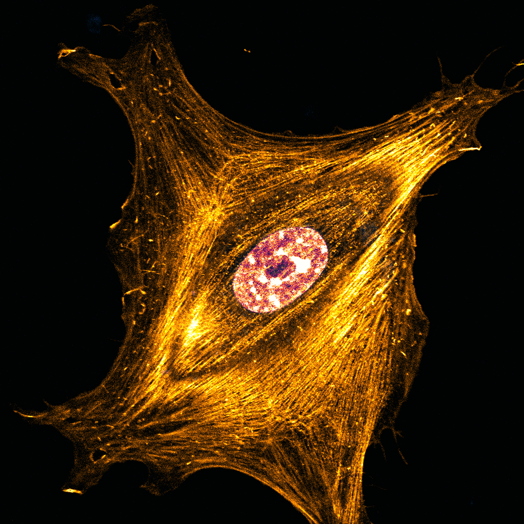
This image may resemble a distant galaxy captured by a space telescope, but the scene is far closer to home. It shows a single skin cell just 10 micrometres across – around one-tenth the width of a human hair.
The cell is growing on a stiff, gel-like material in the laboratory, where it experiences physical forces similar to those found in damaged tissue. The bright, fiery strands stretching across the cell form part of its internal support system, helping it sense and respond to its environment.
Researchers are investigating whether physical forces in a cell’s environment can trigger a remarkable process known as dedifferentiation, where mature skin cells regain stem cell-like properties. This ability helps wounds heal, but the underlying mechanisms are still poorly understood.
Chronic wounds are linked to an increased risk of skin cancer. By understanding how cells respond to physical forces and change their identity, researchers hope to uncover new clues about the biological connection between wound repair and cancer development.
This image is a reminder that extraordinary beauty exists at every scale, from vast galaxies in deep space to the hidden universe contained within a single cell.
Joint Third Prize
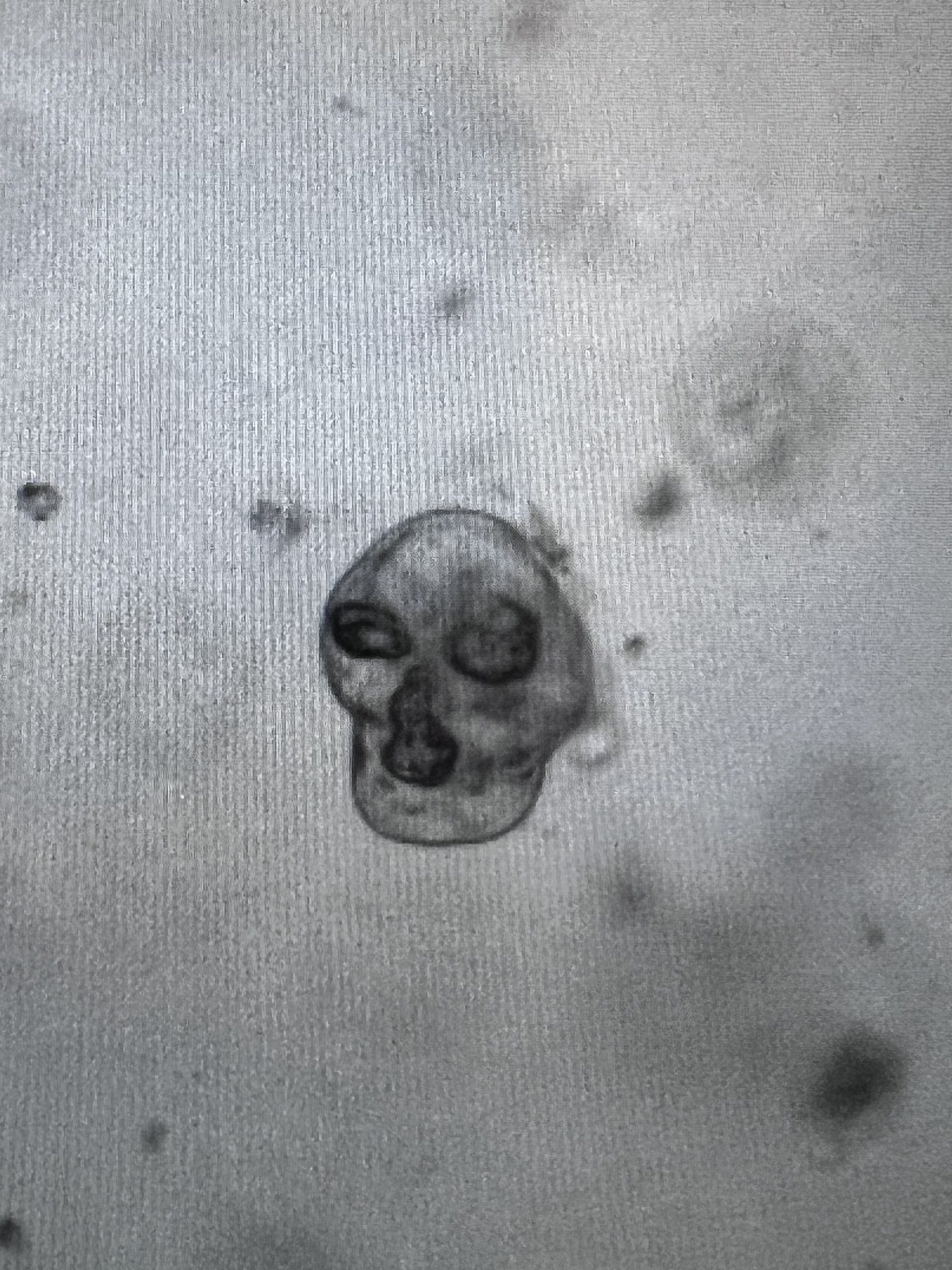
Petra Vlckova (UCL)
CR(C)anium
This image shows a miniature colorectal cancer (CRC) tumour grown in the laboratory. Known as an organoid, it is a 3D model created from a patient’s cancer cells and recreates many of the features of the original tumour.
Its skull-like appearance is a powerful reminder of the fear and uncertainty that can accompany a cancer diagnosis. Yet it also represents hope.
Organoids allow researchers to study cancer in a controlled environment without putting patients at risk. Because they closely resemble real tumours, they can be used to follow how a cancer develops and provide a valuable way to test potential treatments and explore why some therapies work better than others.
In the future, organoids could help doctors personalise treatment by identifying which medicines are most likely to benefit an individual patient before therapy begins.
Capturing both the frightening nature of cancer and the promise of future breakthroughs, this image reflects the determination of researchers working to better understand the disease and develop more effective treatments for patients.
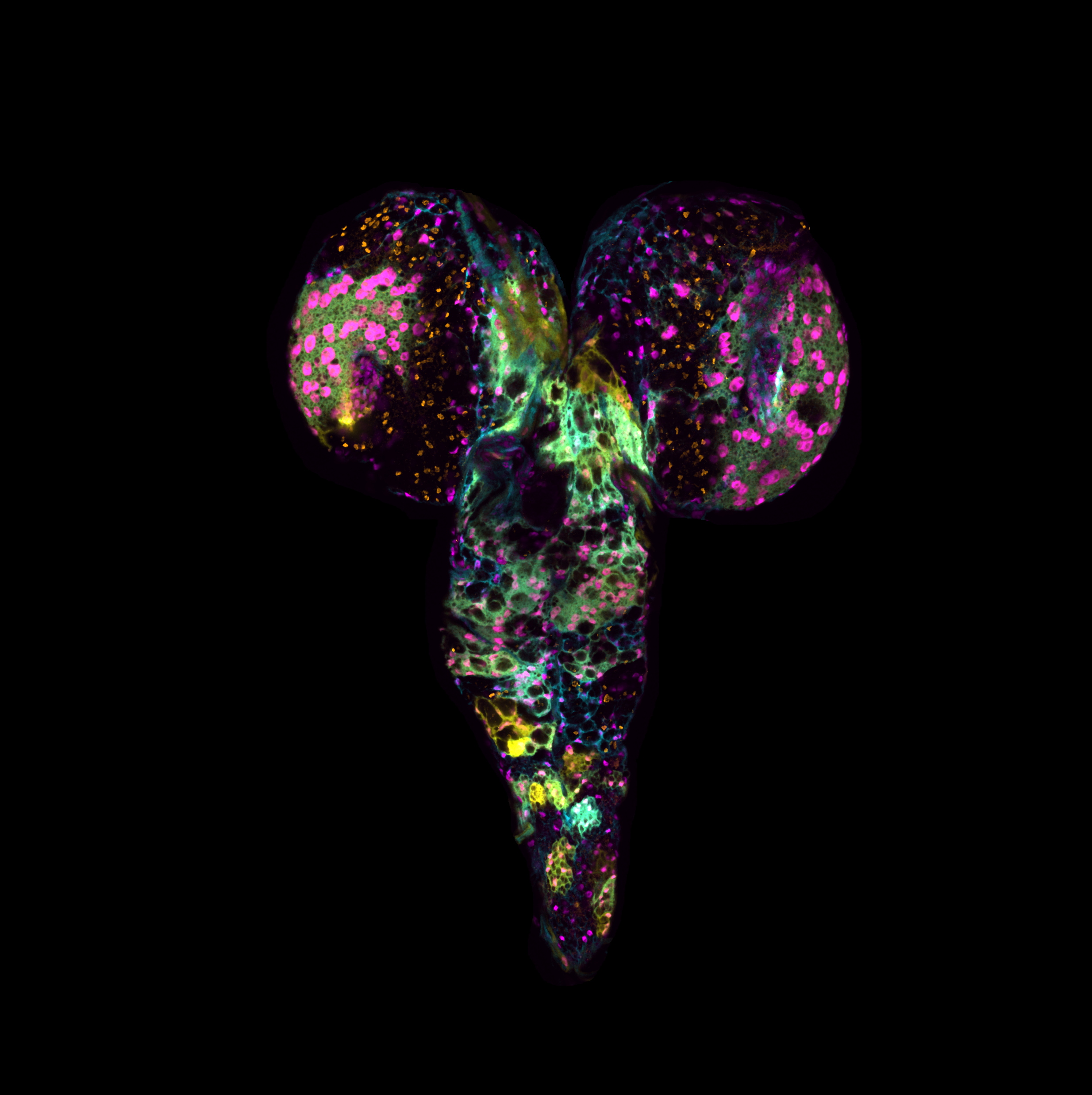
Hannah Burgess (BCI)
Off to a flying start
This colourful image comes from an unexpected ally in the fight against cancer: the fruit fly.
Although tiny, fruit flies share many important biological processes with humans, making them valuable models for studying disease. Here, researchers have used genetic tools to recreate key features of glioblastoma, an extremely aggressive form of brain cancer with limited treatment options.
The different colours highlight cells carrying different cancer-related changes within the tumour model. Rather than acting alone, these cells can work together, helping the cancer grow, adapt and resist treatment.
One of the biggest challenges in glioblastoma research is understanding how different groups of cancer cells communicate and influence each other’s behaviour. By studying these interactions in fruit flies, scientists can explore tumour biology in ways that more closely reflect what happens in patients.
This is particularly important for treatments targeting a protein called epidermal growth factor receptor (EGFR). While EGFR-targeted therapies have shown promise in other cancers, they have largely failed in glioblastoma. Researchers hope that more realistic models like this one will reveal why and help guide the development of more effective treatments for people diagnosed with one of the most difficult cancers to treat.