Image Competition 2025 – Winners
First Prize
Elly Tyler (BCI)
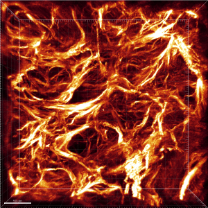
Where Collagen Burns and Immunity Falters
In this breast tumour tissue, collagen fibres in the extracellular matrix blaze like wildfire.
This striking image was created without staining, by using a second harmonic generation multi-photon microscope that emits two photons of light to cause collagen to “glow” and reveal its structure. Collagen is an important part of the scaffolding of tissues known as the extracellular matrix. The structure and density of this matrix affects the ability of our body’s immune cells to move around and engage with tumour cells.
In triple negative breast cancer, which is the most challenging breast cancer type to treat, the collagen matrix is especially dense. This makes it harder for T cells – part of the body’s immune system – to move in and attack the cancer. The cancer is also known to make changes to proteins in the extracellular matrix, making an immune response even more difficult.
By using a process called decellularisation, we can strip out cells and leave only the extracellular matrix, which can then be studied alone, as shown here. Understanding how the architecture of the extracellular matrix changes when cancer is present could pave the way to the development of new treatments. These would aim to boost immune responses to breast cancer and other cancer types where tumours are made up of a solid mass of cells.
Second Prize
Beatrice Malacrida and Florian Laforêts (BCI)
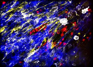
Chaos or Cancer?
“Paint thrown in anger, leaving stark strips of yellows and blue, like tears in reality… or maybe something else?”
This image shows a lab-grown ovarian tumour model, created to reproduce both the chaos and structure of the disease and help scientists to better understand how a tumour interacts with its surrounding microenvironment.
The picture shows the extracellular matrix (yellow), the scaffolding of human tissue. In blue, we see fibroblasts – cells making up connective tissue – and mesothelial cells, which provide a slippery layer between organs. Macrophages (white) are cells of the immune system that prowl the tumour microenvironment in search of cancer cells (red), in order to kill and eat them.
However, cancer can corrupt macrophages so they stop recognising it as a threat, and even ultimately help it spread. By researching the mechanisms enabling this, scientists hope to find new treatment options for ovarian cancer, which is currently especially challenging to treat.
This image was made using a spinning disc fluorescence microscope, funded by the CRUK City of London Centre at the Barts Cancer Institute. This technology supplies the oxygen and carbon dioxide needed by the tumour and keeps it at a consistent body temperature while a time lapse film is captured, showing the cells moving within.
Third Prize
Joshua Daoud (BCI)
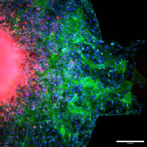
Symbiotic Entanglement
Glioblastoma is a brain cancer that is commonly treated with surgery, chemotherapy and radiotherapy. Sadly, the cancer is always expected to recur, as it is extremely difficult to remove all the cells around the edge of the tumour, and these cells are also most likely to be resistant to treatment.
This image shows glioblastoma (in red) as it invades and becomes entangled with healthy tissue (green) made up of astrocytes, the star-shaped cells that support the neurones within the brain. There is a highly complex relationship between glioblastoma and astrocytes, with some evidence suggesting that astrocytes can aid tumour invasion. However, our understanding of this mechanism is currently limited.
This relationship has been recreated within a microfluidic model – also known as an “organ on a chip” – to allow researchers to study the complex interactions between cells, while reducing the need for animals to be used in research.
By creating a 3D model containing different cell types, researchers can examine cancer cells invading healthy tissue, providing more insight into their behaviour. By understanding these interactions, it is hoped that scientists can find new avenues of treatment for glioblastoma.
Highly Commended
Daire Hanna (BCI)
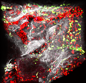
Breaking Barriers: Overcoming Immunosuppression in Pancreatic Cancer
This image shows a lab-grown 3D model of pancreatic cancer. It was captured by a high-resolution multiphoton microscope, which uses multiple photons of light to produce sharp and detailed images.
The model was built using pancreatic tumour tissue from mice, stripped of living cells to leave the dense extracellular matrix behind. This web-like structure (shown in white) is made up of collagen and elastin.
In this image, three key cell types have been added back into the scaffold of the extracellular matrix. These are cancer cells (red); fibroblasts, which are key cells for producing matrix (green); and finally CAR T-cells (pink). These are immune cells that have been engineered to target pancreatic cancer.
Immunotherapy – treatment to help the immune system attack cancer – has had little success in pancreatic cancer. This is mainly because immune cells struggle to reach the cancer cells through the dense barrier of the extracellular matrix. This model recreates the hostile tumour environment in the lab, allowing researchers to test new drug combinations that aim to remodel the tumour and make it more accessible to immune cells.
Ultimately, the goal is to find ways to improve the effectiveness of immunotherapy for people with pancreatic cancer.
Eitan Mirvis (KCL)
Finding gold in the middle of the rainbow
“In Irish mythology, gold lies at the end of the rainbow. But in our lab, we find it in the middle.”
CAR-T therapy is a ground-breaking treatment that targets cancer more effectively by reprogramming a patient’s T cells – part of the immune system.
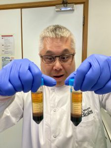
This image shows Dr Le Anh Luong, senior research assistant in Dr Reuben Benjamin’s Cellular Immunotherapy Lab at King’s College London. Dr Luong holds blood samples that have been separated into different components. This allows scientists to collect T cells, the “police officers” of the immune system, from the blood. The T cells are white blood cells found in the very thin “buffy coat” that lies between the plasma (orange) and the Histopaque, the straw-coloured layer that is added to separate out thinner and thicker parts of the blood when spun in a centrifuge. At the bottom of the tube, we find red blood cells and granulocytes, another type of white blood cells.
Once collected, the T cells are genetically engineered to identify and kill cancer cells, before being returned to the patient.
In the photograph, Dr Luong marvels at the ground-breaking potential of T cells to cure blood cancers. The image is full of personality, showing the friendly, vibrant atmosphere in the lab.
Carmen Rodriguez (UCL)
Osteoclasts at work inside a bone tumour
Osteosarcoma is a cancer of the bone. It is characterised by a harmful interaction between the tumour and osteoclasts, specialist cells that play an important role in remodelling healthy bone, allowing it to grow by breaking it down and reabsorbing it.
In cancer, the tumour exploits this process, creating more space for itself. In turn, osteoclasts send signals that encourage the tumour to grow. This ‘vicious cycle of the bone’ creates a tumour-friendly environment that supports cancer growth and spread.
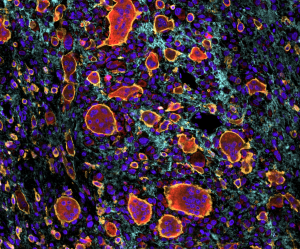
This image shows immune cells (yellow) inside osteosarcoma, surrounded by collagen I (blue), the main component of the bone. Among them are the osteoclasts (red), which have multiple cell nuclei, the control centres of the cell (purple).
This image was captured using the MACSima multiplex immunofluorescence platform, which allows scientists to carry out multiple rounds of staining and imaging. This means they can detect many protein markers within a single cell, providing valuable insights.
Capturing images like this one helps researchers to explore the multiple barriers that prevent immune cells from effectively attacking bone tumours. They hope to use this understanding to develop more effective next generation CAR T cell therapies, engineering the body’s own immune cells to find and attack osteosarcoma.
Nick Willoughby (KCL)
Explaining how blood samples lead to cancer breakthroughs
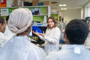
In this photo, PhD student Esme Carpenter is explaining to cancer patients how their precious blood samples are used to explore the tumour microenvironment. The tumour microenvironment includes the surrounding cells, blood vessels, and immune cells that interact with cancer.
This photograph was taken during an engagement day for Project SAMBAI (Social, Ancestry, Molecular and Biological Analysis of Inequalities). This study is part of Cancer Grand Challenges, a global initiative tackling the toughest challenges in cancer research. Project SAMBAI aims to improve our understanding of poor cancer outcomes in under-served populations of African descent.
During the engagement day, patient contributors were updated on the project and invited to give their thoughts. Here, a group is being shown the laboratory equipment used to investigate their samples.
The image was taken using a Sony A7 III. By attaching the flash to the top of the camera, the photographer bounces light off the ceiling of the laboratory to produce a more diffuse, less harsh effect. Working with the flash makes it difficult to “cheat” by using burst mode to take multiple photos in quick succession, so this expressive image is the result of skilful – and lucky – timing.
