Integrated Infrastructure Core Platforms
Shared state-of-the-art technical infrastructure is an important aspect of the Centre’s strategy, enhancing the research of more than 100 groups across its multiple sites.
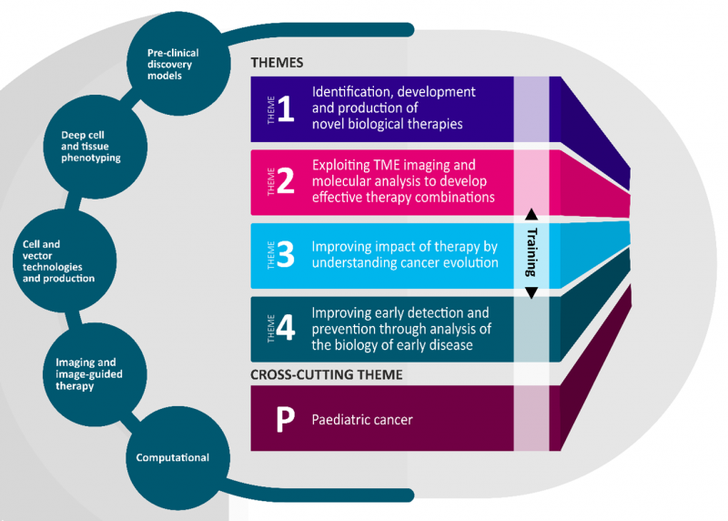
Specialist shared cores give the Centre five diverse capabilities, supporting discovery research in pre-clinical and human tissue models through to the provision of cell-based therapies and imaging tools for clinical trial.

The Centre also contributes to the support of more ‘routine’ research infrastructure distributed across the Centre sites, including flow cytometry, microscopy and imaging, proteomics, genomics, bioinformatics, pathology, organoid, cell engineering, and GCLP. This support is vital for the success of CRUK-funded programmes at the Centre.
UCL Cancer Institute https://www.ucl.ac.uk/cancer/research/cancer-institute-translational-technology-platforms
- A 3D bioengineering specialist at the Barts Cancer Institute to identify collaboration opportunities with other Centre labs that are using these technologies, develop SOPs for multi-cell 3D human tumour models that can be used throughout the Centre and identify the need for new techniques. See more details about Human Tumour Microenvironment Models here. Contact: Mina Mincheva: m.mincheva@qmul.ac.uk
- A Tcom PDX/iGEMM specialist at the UCL Cancer Institute, to support development of facilities at Clare Hall, identify pilot project opportunities with the Crick and other partners, and generate relevant SOPs. See more details about Patient-Derived immune Xenograft Core here. Contact: Mansi Shah: mansi.shah@ucl.ac.uk
- ZellScannerONE™ chipcytometry to enable cutting edge, multiparametric analysis of cell-to-cell variations within cell populations. A technician is available to run the chipcytometry system. The scanner is located at Barts Cancer Institute. See more details about the Zellscanner One Imaging Platform here. Contact: Joseph Hartlebury j.hartlebury@qmul.ac.uk
- A LSM 880 with Airyscan Fast confocal microscope at Barts Cancer Institute. See more details about the Zeiss LSM 880 Confocal Microscope with Airyscan Fast here. Contact: Linda Hammond l.j.hammond@qmul.ac.uk | +44 (0)20 7882 5132
- A small cabinet irradiator is available as part of a preclinical irradiation platform for focal delivery of radiotherapy, to model combination therapy with radiotherapy at UCL. See more details about the Preclinical Radiotherapy core here. Contact: Rebecca Carter rebecca.carter@ucl.ac.uk
The Centre hosts 2 single cell specialists at the UCL Cancer Institute. The post will build links with the TME models team, identify data storage/analysis needs, generate protocols, and initiate pilot projects primarily aimed at single cell analysis of the TME in cancer samples. See more details about the Single Cell Genomics Facility here. Contact: Imran Uddin imran.uddin@ucl.ac.uk | Gordon Beattie g.beattie@ucl.ac.uk
Equipment available for the Cell and Vector Manufacturing Core
-
Four isolators for vector manufacture to support development of cell therapies and clinical translation, with the aim of minimising immune-related toxicities in adult and paediatric cancer at KCL
Expertise available for the Cell and Vector Manufacturing Core
-
A GMP cell production specialist is located in the Centre for Cell, Gene & Tissue Therapeutics at the Royal Free Hospital. The specialist will initiate SOPs for pilot projects, primarily of CAR, TCR and TIL therapies in adult and paediatric cancers, as an initial enhancement of GMP manufacturing capacity.
-
An additional 1 GMP cell production specialist will be available at the Centre for Cell, Gene & Tissue Therapeutics at the Royal Free Hospital. Contact: Amaia Cadinanos Garai a.garai@ucl.ac.uk.
-
Two GMP specialists will be at the Zayed Centre for Research into Rare Disease in Children. Contact: Louisa Green louisa.m.green@ucl.ac.uk.
See more details about the Cell and Vector Manufacturing Core here.
The Imaging core primarily supports the Cross Disciplinary Approaches Enhancing Biotherapeutics programme in the CRUK City of London Centre.
This core utilises a diverse range of imaging modalities and inter-disciplinary approaches to explore how best to develop and use biological therapies. This includes imaging the distribution of biological therapies and imaging changes in tumour volume, perfusion, vascular structure, and tumour mechanics. Imaging modalities include MRI, PET, SPECT, microCT, and two-photon microscopy.
Equipment available at the Imaging and Image-Guided Therapy Core
- A clinical spec 3T MRI scanner is available into the research environment at UCL. This equipment will enable monitoring tumour response to biotherapeutics and conventional therapies, and accelerate the translation of new protocols from the lab to the clinic.
- A pre-clinical CT-guided irradiator at UCL. Contact: Rebecca Carter rebecca.carter@ucl.ac.uk
A centralised High Performance Computing (HPC) Cluster storage and processing units, supported by 2 HPC Technology Staff is embedded within the UCL Department of Computing. Additionally, a specialist for data integration is based at UCL. See more information on the CoL Centre HPC Cluster here.
